Tissue Characterisation Consulting
Ultrasound Imager Quality Control and...
...3D Imaging and 3D Image Processing
Quality Control System for Ultrasound Imager (details)
Data acquisition
The sufficient phantom data can be collected with acquisition system using the frame grabber. Frame grabber transfer the video image of ultrasound equipment to the personal computer creating the 3D phantom image.
Frame grabberBy 3D imager the 3D-image of the phantom can be taken directly from imager. With 2D imager the free hand scan is possible but the scan adapter deliver the geometry of phantom structure more exact.
Picture show special fastening system (platform) for vaginal probe. Different fastening system can be used for large varieties of probes. The platform will be hold onto phantom directly by 4 screws (bottom).3D image is sequence of B-images. With loss less packing and compressing the image data, the smallest possible file formats are achieved.
Large amount of data will be stored at external HD disk in order to keep the special date separately from other content at the working disk of the personal computer.
External 40GB HD-diskData processing
…produce more different images for quantitative and visual impression of probes quality.
The first processing step is gray level calculation in tree perpendicular planes, which show the visual impression of azimuthal and elevational resolution. The same planes show the saturation inside the gray level window if it take place.The gray values mustn’t reach the saturation. Saturated images deliver wrong SNR values.
Calculated SNR give the information about the ability of cyst detection for given size and depth. The functional range is defined with SNR greater then 2.5 (SNR>2.5).If the SNR diagram is combined with C-images, the visual control of SNR function will be performed showing the ability of detection limits for artificial cyst of the given size.
At current time doesn’t exist the general automated utilization of ultrasound medical images. The visual way is still the mostly used method and practice. The visual control of phantom image is confirmation of calculated parameters on order to avoid the possible misinterpretation.
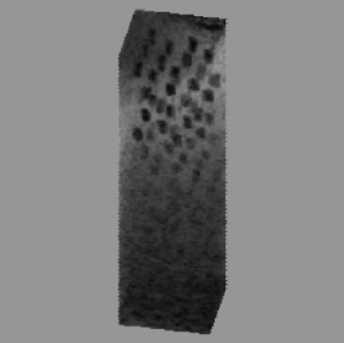
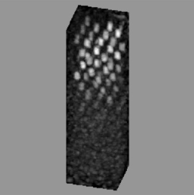
SNR diagram and visual check on C-imagesIn order to achieve the spatial impression of phantom structure, the rendering images can be shown:
Projection of phantom image with artificial cysts presentation..jpg)
.jpg)
.jpg)


 TCC
TCC